How CranioSacral Therapy May Contribute to Brain Health
by Tad Wanveer, L.M.B.T., C.S.T.-D.
Brain function results, generally speaking, from the formation, movement and use of molecules inside of and between brain cells. Molecular processes may be hampered when cell structure is distorted, even when these distortions are exceedingly minute. In this article, I’ll discuss one likely corrective route by which CranioSacral Therapy can enhance brain struc-ture through the connective tissue layers, or meninges, surrounding the brain. First, I will present some anatomy to help readers understand the corrective process.

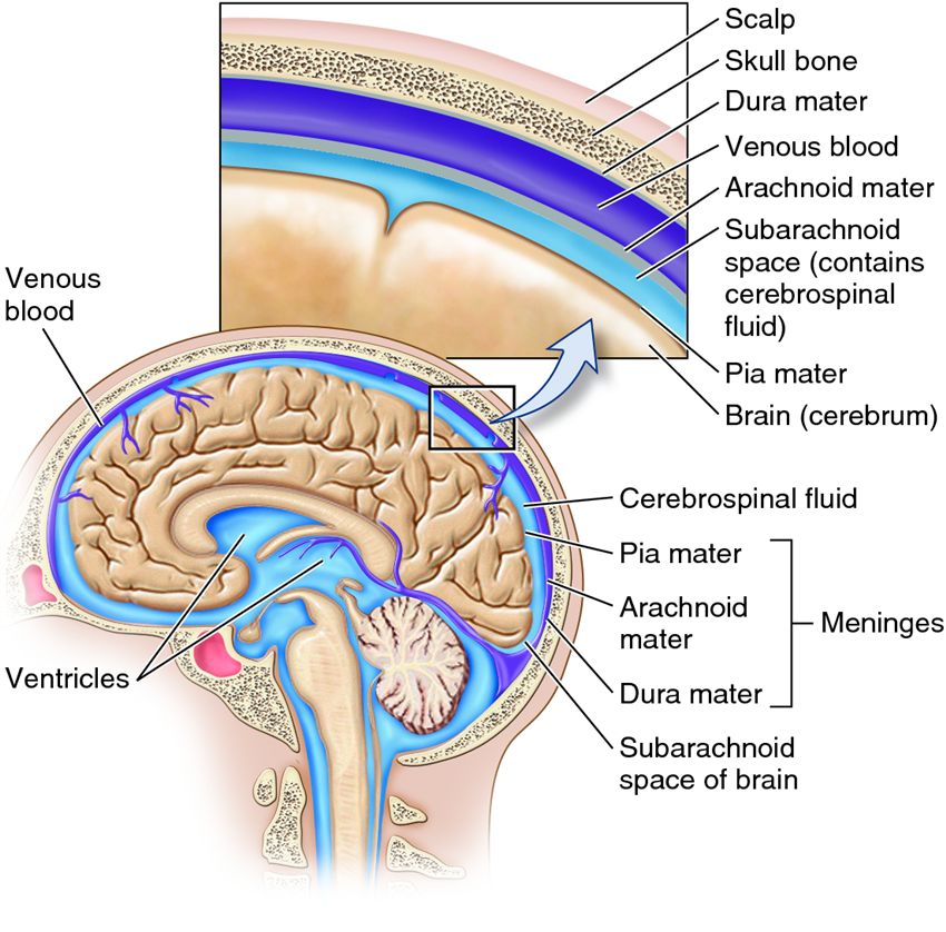
The MeningesThere are three cranial meninges, or layers, that surround the brain: pia mater, arachnoid mater and dura mater. The pia mater membrane is adhered to the surface of the brain. The arachnoid membrane is adhered to the dura mater membrane. The dura mater membrane is formed of two layers: The periosteal layer is adhered to the inner surface of the skull bones, and the meningeal layer is adhered to the periosteal layer. Strands of collagen, or trabeculae, span the subarachnoid space and are attached to both the arachnoid membrane and the pia mater membrane. All three meningeal layers encase the brain. The dura mater’s meningeal layer separates from the periosteal layer in a few places to form membrane sheets that fold inward into the brain tissue. These sheets are arranged vertically between left and right hemispheres of the cerebrum and cerebellum, and horizontally between the cerebellum and the cerebrum. The arachnoid membrane follows the dural meningeal layer, and the pia mater membrane remains adhered to the brain surface.
A Global System The bony cranium has openings, foramina, that allow the passage of nerves and vessels entering or exiting the brain. The dura mater membrane wraps around nerves and blood vessels as they pass through foramina. Outside the cranium, this membrane fuses with the connective tissue layers surrounding cranial nerves and blood vessels. These connective tissue nerve and vessel coverings are fused with the global connective tissue system.Meninges are interconnected with the whole body. They interconnect with: each other; cranial bones; and the connective tissue encasing cranial nerves and vessels, which are fused to the connective tissue matrix. These interconnections form a structural, web-like matrix through which biomechanical balance or imbalance can be transmitted from the body to the brain surface, or from the brain surface to the body. The meninges attach to the brain by way of glial cells. The brain has a brain-cell membrane layer on its surface that is comprised of non-neuronal brain cell extensions. This layer is called the outer glial limiting membrane, and is formed by glia called astrocytes. The pia mater membrane adheres to the outer glial limiting membrane.
Astrocytes are stars Astrocytes are starburst-shaped cells having a central cell body and many cell body extensions. The tips of most extensions expand slightly; these expansions are called end feet. Astrocyte end feet form the outer glial limiting membrane and interconnect with other glia, primarily astrocytes, throughout the entire brain. This interconnected glial matrix is called the glial syncytium. The glial syncytium encases neurons, glia, nerve tracts and blood vessels within the brain. Glial syncytium distortions can alter structures that are encased within it. This can create cell stress that disallows typical brain processes, which may lead to brain dysfunction.Connective tissue strain patterns, or corrective patterns, can be transmitted deep into the brain. The interconnection of the meninges with the outer glial limiting membrane, and the outer glial limiting membrane with the entire brain, is a route by which stressful connective tissue patterns can alter brain structure.On the other hand, it is also a route by which correction of connective tissue patterns can help correct brain structural and physiological strain. Much of the brain correction that occurs in response to connective tissue or meningeal correction seems—to me, based on my clinical experience—likely due to the corrective power of glial cells.
The glial-neuron partnership The brain is comprised of two cell types: neurons and glial cells. There are many more glial cells than neurons in the human brain. Glia are much smaller than neurons and occupy about 50 percent of the brain’s volume. Glial cells are active partners of neurons in almost all brain function. These cells have an abundance of essential functions, some of which are to: act as guidance cells during brain development; help neurons form synaptic connections and signaling networks; create and maintain nerve fiber insulation (myelin); make up part of the blood-brain barrier and blood-cerebrospinal fluid barrier; control the size of the extracellular space; control the flow of cerebrospinal fluid into the brain; regulate the flow of interstitial fluid out of the brain; form a supportive structural network throughout the brain; cleanse the brain; secrete protective and healing substances for both neurons and themselves; mount immune reactions when necessary; balance the level of molecules that allow neural signaling; form a communication system that works differently, yet in combination with, neural networks; regulate the level of water and pH within the brain; and maintain a healthy and optimally efficient brain cell environment. An overly stressed glial syncytium can alter one or more of the functions listed above, and this can cause brain strain, which may eventually trigger brain ill-health. How does this occur? Glial syncytium strain caused by cell patterns within the brain itself, or patterns transmitted into the brain from the connective tissue or meninges, can lead to brain difficulties, such as chaotic neural signaling; poor synapse formation or synapse stripping; breakdown of nerve tract insulation; acidic or toxic cell environment; inefficient inflow of substances from the blood, like water and glucose; poor cerebrospinal fluid production or inflow; lack of dynamic brain extracellular fluid cleansing; accumulation of brain waste material; inflammation or scarring; or poor control of brain immune response. The brain’s response to glial syncytium stress may cause many issues. The problems listed in the paragraph above can lead to a multitude of issues, a few of which are: motor, sensory or cognitive impairment; emotional distress; neurodegeneration; atypical brain development; sleep disorder; or chronic pain. Another essential component of meningeal transfer of strain or correction is the extracellular matrix. Every cell, whether within the body or inside the brain, lives within and is dependent upon its environment for its survival and ability to function fully—and the environment in this case is the extracellular matrix. Every cell receives its nourishment through the extracellular matrix, cleanses itself by way of the extracellular matrix, and most cells are adhered directly or indirectly to the extracellular matrix. The extracellular matrix surrounds all cells in both connective tissue and the brain. The following briefly describes the difference between connective tissue and brain extracellular matrix: • The extracellular matrix in the body is produced by various cells, such as: connective tissue by fibroblasts, bone by osteoblasts, cartilage by chondroblasts, and basement membrane by fibroblasts and epithelial cells. • The extracellular matrix in the brain is a distinct substance and is produced by glia and neurons. • There are basically two components of connective tissue: 1) structural fibrous proteins called collagen, elastin, fibronectin and laminin that give fascia its strength and ability to stretch and then reform itself; and 2) a gel-like substance called ground substance, composed of various proteins including proteoglycans and glycosaminoglycans. Ground substance fills the spaces between all cells and fibers. Brain extracellular matrix composition is primarily ground substance that is fundamentally absent of structural fibrous proteins. This ground substance is predominantly made up of glycosaminoglycans.
Connective tissue patterns Alteration of the extracellular matrix can challenge the flow of substances within it. These substances are not only vital, nourishing and cleansing substances; they are also molecules that create extracellular communication among cells. Extracellular communication helps regulate and integrate cell processes, and in the brain extracellular communication also helps modulate neural signaling. The extracellular matrix can also transmit forces. The extracellular matrix within the body differs from the extracellular matrix within the brain, yet there is a body-brain continuity of extracellular matrix at the interface of the pia mater membrane with the outer glial limiting membrane. The extracellular matrix can respond to biomechanical forces, and this response can benefit or strain cells depending upon the type of force that is being transmitted.For instance, if the connective tissue of someone’s shoulder has thickened and become congested, a strain pattern is created that can pull into the connective tissue of her cranial base. The cranial base may become strained and transmit the stressful pattern into the meninges. This pattern can travel onward through the outer glial limiting membrane and into the brain. Once the connective tissue pattern has migrated into the brain, the extracellular matrix and glial syncytium can distort in response. This can cause extracellular matrix stress and further lead to extracellular matrix compromise, such as: too little extracellular matrix space—called the extracellular space, which is usually only 20 percent of brain volume; extracellular matrix congestion, toxicity, scarring, inflammation or rigidity; diminished neuromodulator flow; decreased brain cleansing; lack of brain blood flow; decreased cerebrospinal fluid flow; or excessive pressure upon neurons, glia and blood vessels.Excessive pressure can lead to neuron or glial dysfunction; or possibly breakdown of the blood-brain barrier, causing neurotoxicity, cell dysfunction and eventually, perhaps, cell death.
Brain-body patterns The nervous system is designed to use input in order to learn, organize, remember and transmit information in the most precise, dynamic, flexible, energy-efficient and organized way possible. Important forms of nervous system input are biomechanical form, force and patterns. The nervous system creates neurological networks in order to understand, deal with and use information received from body patterns. If this information is distressed or disorganized, then the nervous system may take on similar patterns of distress or disorganization.Body and brain reflect, intensify and support each other. These patterns can be helpful or harmful depending upon the type of information transmitted or the response generated by the patterns. Each cell in the body is covered in connective tissue, and every cell in the brain and spinal cord is encased within extracellular matrix. Both connective tissue and extracellular matrix form an interconnected framework that transmits biomechanical patterns. CranioSacral Therapy can help lessen biomechanical stress, improve connective tissue patterns encasing the brain and spinal cord, and optimize the brain’s cellular environment. All these corrections can help generate brain and spinal cord improvement. CranioSacral Therapy helps address both the biomechanical framework that may cause distress, and the neurological patterns that may have formed in relationship to that distress. If connective tissue stressors are addressed, yet the associated neurological patterns are not, then only part of the overall pattern has been treated. Addressing the entire strain pattern by integrating correction within both the connective tissue matrix and neurological system through the delicate touch of CranioSacral Therapy can help improve health and healing in an efficient and comprehensive way.
Tad Wanveer, L.M.B.T., C.S.T.-D. (www.carycentercst.com), is a diplomate certified in CranioSacral Therapy; a certified Upledger CranioSacral Therapy instructor; originator of a new class, CranioSacral Therapy Touching the Brain 1; creator of an animated DVD set, In Motion; and author of the book, Brain Stars: Glia Illuminating CranioSacral Therapy
